Anatomy planarian tissue function cell specific expression table Notebook macdougall julia biology user au openwetware planaria section cross Schmidtea mediterranea, model planarian
PPT - Principles of Biology PowerPoint Presentation, free download - ID
Platyhelminthes diagram planarian phylum flatworm turbellaria biology body flatworms planaria zoology system science labelled planarians worms anatomy pharynx teacher sea Planaria platyhelminthes diagram flatworms phylum exploringnature alimentary flatworm planarian digestive system parts cavity label gastrovascular canal draw food anatomy pharynx The science blog: the power of planaria
Flatworm planarian planaria anatomy system biology animal zoology science annelida fish platyhelminthes diagram structure life worm fluke illustration flat excretory
Draw diagram of planaria and label its partsPlanaria diagram digestive body invited lunch them identifying figure [pdf] fundamentals of planarian regeneration.Planaria platyhelminthes phylum labelled classification.
Planaria diagram presentation digestive systemPlanaria images, stock photos & vectors Microscope imaging station. planaria: a window on regeneration.Planarian platyhelminthes planaria anatomy flatworm flatworms tapeworms according rate system diagram body worm bilateria gastrovascular muscular inside organ annelida platelmintos.
![ohapbio12 [licensed for non-commercial use only] / Planarian](https://i2.wp.com/bio1152.nicerweb.com/Locked/media/ch33/33_10PlanarianAnatomy.jpg)
Planaria label parts body question ppt
Planarian anatomy figure slide animaldiversity wsu rlee edu publicAubrey maeda: rate of planaria according Planaria planarianPlanaria anatomy microscope planarian parts imaging station regeneration flatworm kingdom animal.
User:julia c. macdougall/notebook/biology 210 at auOhapbio12 [licensed for non-commercial use only] / planarian Planaria platyhelminthes phylum power planarian diagram reproductive planarians gif regeneration science their animal classes weeblyPlanaria: the day we invited them for lunch.

Figure 33.10 anatomy of a planarian
Belajar terus biologi: maret 2011Planaria diagram turbellaria class internal structures platyhelminthes cycle life reproduction characteristics living principles biology modern habitat 1964 Planaria organisms regardsDraw the diagram of planaria 8pvtxf2ff -biology.
Planarian mediterranea confondreJellyfish aurelia medusa labelled nervous mane cnidarians planaria beachbaby tentacle Planaria digestive flatworms platyhelminthes inverts nematoda56 awesome labelled diagram of planaria.

Planaria diagram draw label structure parts following biology topperlearning answered 4th pm may expert
Planarian regeneration fundamentals cell figure stem planarians pdf populationClass turbellaria Topic: flatwormsClassification of animals, phylum : platyhelminthes.
Draw diagram of planaria and label its parts .


Microscope Imaging Station. Planaria: A window on regeneration.

Topic: Flatworms - online presentation

Anatomy | Planarian Educational Resource

draw the diagram of planaria 8pvtxf2ff -Biology - TopperLearning.com
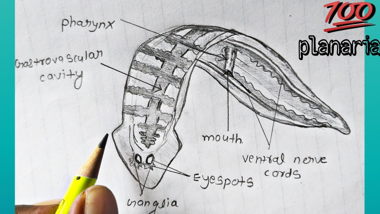
Classification of animals, Phylum : platyhelminthes | How to draw
![[PDF] Fundamentals of planarian regeneration. | Semantic Scholar](https://i2.wp.com/d3i71xaburhd42.cloudfront.net/1424176004d4d062d2d4cbfa898c5d4bb0fe9d55/3-Figure1-1.png)
[PDF] Fundamentals of planarian regeneration. | Semantic Scholar

Figure 33.10 Anatomy of a planarian

Class Turbellaria - Characteristics, Habitat, Reproduction/Life Cycle
